Home
/ Cross Section Of A Compact Bone - Pin by Danielle Papas on Nursing Anatomy & Physiology ... : These are abundant and characteristic of compact bone.
Cross Section Of A Compact Bone - Pin by Danielle Papas on Nursing Anatomy & Physiology ... : These are abundant and characteristic of compact bone.
Cross Section Of A Compact Bone - Pin by Danielle Papas on Nursing Anatomy & Physiology ... : These are abundant and characteristic of compact bone.. The spongy and compact bone tissue in the cross section of a skull bone. Osteocyte processes lie in tiny canals (canaliculi) in the bone matrix. Cross section of compact bone. As the names suggest compact bone looks compact and the spongy bone looks like sponges. The osteon consists of a central canal called the osteonic (haversian) canal, which is surrounded by concentric rings (lamellae) of matrix.
Select different colors for the. The osteon consists of a central canal called the osteonic (haversian) canal, which is surrounded by concentric rings (lamellae) of matrix. Cross section of compact bone. To know the structures of a synovial joint and a symphysis joint (intervertebral disc). Bone decalcification is the removal of the mineral component using an acid, leaving the bone soft and easy to cut.
A and P Lab section 8 flashcards | Quizlet from o.quizlet.com From wikimedia commons, the free media repository. Spongy bone is the osseous tissue, which fills the interior cavity of bones, consisting of mineralized bars called trabeculae. As the names suggest compact bone looks compact and the spongy bone looks like sponges. This is a cross section through decalcified bone. Most bones contain both compact and spongy bone. A cross section of a compact bone shows concentric circles called lamellae. There are two ways to study bone histology. (micrograph provided by the regents of university of michigan.
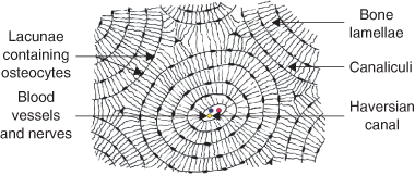
A diagrammatic view of a cross section of bone.
The remainder is spongelike cancellous bone. Cross section of compact bone. The two layers of compact bone and the interior spongy bone work together to protect the internal organs. Structures and bone areas in column b, and use them to color the coding. Their course follows the main axis of long bone. (micrograph provided by the regents of university of michigan. In the center of each osteon is the central canal, a space that houses blood vessels and nerves that supply bone. Don't assume that the cross sectional area is the same no matter where you cut. There are two ways to study bone histology. Canaliculi allow the passage of interstitial fluid between the central canal and the lacunae housing osteocytes. Bone must be decalcified (by exposure to strong acids) so it can be cut into thin sections. As the names suggest compact bone looks compact and the spongy bone looks like sponges. Remodeling allows the body to fix damaged sections, reshape the skeleton during growth, and regulate calcium levels.
Their course follows the main axis of long bone. These are mostly compacted bone with little marrow and include most of the bones in the limbs. An estimated 10 percent of an adult's skeleton is replaced each year. To recognise bone and understand its structure and to understand the processes by which bone can be formed. In a cross section of a bone we can see two types of bone tissue:
1 Oral embryology, histology and anatomy | Pocket Dentistry from pocketdentistry.com Select different colors for the. Spongy bone and compact bone. A diagrammatic view of a cross section of bone. The osteon consists of a central canal called the osteonic (haversian) canal, which is surrounded by concentric rings (lamellae) of matrix. Cross section of a femur bone showing the anatomical structure including cancellous bone and marrow. This is a cross section through decalcified bone. Dry bone is cut and polished before mounting on a slide. To know the architecture of compact and spongy (cancellous) bone.
(b) in this micrograph of the osteon, you can clearly see the concentric lamellae and central canals.
As the names suggest compact bone looks compact and the spongy bone looks like sponges. These are abundant and characteristic of compact bone. Spongy bone and compact bone. Compact bone is very different from the other tissues you have seen. Magnification view of compact bone tissue. The two layers of compact bone and the interior spongy bone work together to protect the internal organs. To know the structures of a synovial joint and a symphysis joint (intervertebral disc). Dry bone is cut and polished before mounting on a slide. To recognise bone and understand its structure and to understand the processes by which bone can be formed. They build the entire picture, improve your understanding, consolidate the information and facilitate recall. Compact bones make up 80 percent of the human skeleton; (b) in this micrograph of the osteon, you can clearly see the concentric lamellae and central canals. To know the architecture of compact and spongy (cancellous) bone.
The remaining material is mostly collagen. In the center of each osteon is the central canal, a space that houses blood vessels and nerves that supply bone. A cross section of a compact bone shows concentric circles called lamellae. A central tube called a haversian canal typically runs in the same path as the length of the bone. Their course follows the main axis of long bone.
Skeletal System (2/28-3/4) - Nutritional Sciences 3410 ... from s3.amazonaws.com The two layers of compact bone and the interior spongy bone work together to protect the internal organs. A cross section of a human long bone. Spongy bone is the osseous tissue, which fills the interior cavity of bones, consisting of mineralized bars called trabeculae. Spongy bone and compact bone. A bone is a rigid organ that constitutes part of the vertebrate skeleton in animals. A central tube called a haversian canal typically runs in the same path as the length of the bone. The remaining material is mostly collagen. An estimated 10 percent of an adult's skeleton is replaced each year.
These are mostly compacted bone with little marrow and include most of the bones in the limbs.
To know the architecture of compact and spongy (cancellous) bone. This is a cross section through decalcified bone. Magnification view of compact bone tissue. A bone is a rigid organ that constitutes part of the vertebrate skeleton in animals. Spongy bone and compact bone. Between the rings of matrix, the bone cells (osteocytes) are located in spaces called lacunae. Hope you enjoy and please. To know the structures of a synovial joint and a symphysis joint (intervertebral disc). Also called cortical bone, the compact variety usually features a haversian system, or cylindrical unit within the structure. In a cross section of a bone we can see two types of bone tissue: Select different colors for the. A central tube called a haversian canal typically runs in the same path as the length of the bone. The connection point for the periosteum.
There are trabeculae in spongy bone which gives its sponge like appearance cross section of a bone. (micrograph provided by the regents of university of michigan.

